

Knee Anatomy
There are five bones that meet at the knee.
The end of the femur (thigh bone) has 2 convex surfaces: the medial (inner side) femoral condyle and the lateral (outer side) femoral condyle. These are covered with smooth, white articular cartilage (also known as ‘hyaline cartilage’) and articulate against the upper end of the tibia (tibial plateau) which is also covered in articular cartilage, to form two separate but contiguous joints, the medial tibiofemoral joint and the lateral tibiofemoral joint. In the central area of the tibial plateau are the anterior and posterior tibial spines. In the central area of the femur is a groove (trochlea) where the patella (kneecap) runs. On the lateral side of the tibia, just below the level of the tibiofemoral joint is the upper end of the smaller bone of the calf, the fibula.

The knee is held together by several tough ligaments.
A ligament is a fibrous tissue that attaches two bones together for stability, as opposed to a tendon that attaches a muscle to a bone to pull on it. On the medial side, the medial collateral ligament (MCL) attaches from a point on the edge of the medial femoral condyle (the epicondyle) and runs down to a more diffuse insertion on the medial side of the tibial plateau. On the lateral side there is a more cord-like tendon running from the lateral condyle to the head of the fibula, the lateral collateral ligament, LCL. The MCL and ACL prevent sideways movement of the knee.
Front to back movements are prevented by 2 ligaments running roughly front to back that cross in the notch between the femoral condyles, namely the anterior cruciate and posterior cruciate ligaments (ACL and PCL)
There are several more minor but important ligaments that are thickenings of the capsule that runs completely around the knee such as the posterolateral corner (PLC), the medial patellofemoral ligament (MPFL)
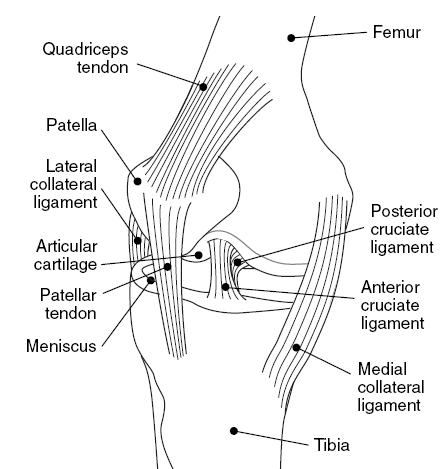
Between the surfaces are two menisci
These are often referred to as ‘meniscal cartilages’ or sometimes just ‘cartilages even though they are made of tough fibrous tissue rather than hyaline cartilage. They are both crescent shape, the lateral meniscus being three quarters of a circle and the medial meniscus nearer two thirds. Both are thick at the outer edge and taper down to the inner surface. Only the outer 20% has a blood supply, so only this part has the potential for healing. Tears of the inner part do not have a blood supply and often will not heal even if repaired perfectly.

There are several muscles around the knee.
The quadriceps have four parts: large muscles from the medial and lateral sides of the femur (vastus medialis and vastus lateralis) and one from the middle of the femur (vastus intermedius). The fourth runs straight up the thigh and across the hip joint to attach to the pelvis (rectus femoris). They merge to form the quadriceps tendon which envelops the patella and continues as the patellar tendon, joining to the tibia at a prominence of bone called the tibial tuberosity. Their main function is to extend (straighten) the knee and stabilise the patella in the trochlea with the tissues on either side, the patellar retinaculum.
Behind the knee are the hamstrings, comprising the biceps femoris laterally attaching from the pelvis to the fibular head, and the semimembranosus and semitendinosus muscles medially from the pelvis to the tibia. They flex (bend) the knee and help extend the hip joint.


There are major nerves and blood vessels at the back of the knee.
The sciatic nerve divides above the knee into the tibial nerve and the common peroneal nerve; the tibial nerve runs with the continuation of the femoral artery and vein (the popliteal artery and vein) into the back of the calf, the common peroneal nerve winds round the neck of the fibula to innervate the muscles at the front and outer side of the calf.
